


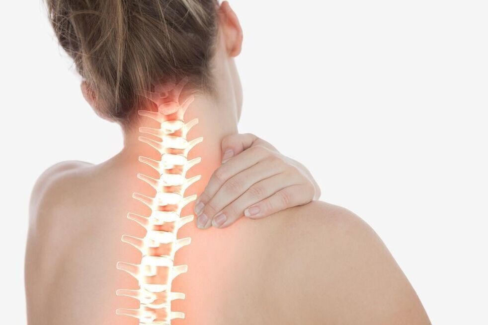
The spine, like any structure that performs a supporting function, inevitably wears out over time. High static and dynamic loads, as well as local overloads of segments of the particularly mobile upper section, lead to a decrease in regenerative capacity and a gradual degeneration of cartilage and nearby muscle-band structures. By the age of 30-35, almost all people show signs of cervical osteochondrosis to a greater or lesser extent. And while it is impossible to stop the irreversible process of biological aging, it can be slowed down completely.
Diagnostics
The following methods are used for the objective assessment of degenerative-dystrophic lesions of the cervical spine and for the detection of degenerative-dystrophic lesions:
- smooth spondylography (X-ray contrast-free examination in frontal, lateral and oblique projections)
- X-ray with functional tests
- MSCT (multilayer computed tomography)
- MRI
- Survey spondylography of the upper spine is a traditional method of radiological diagnosis of cervical osteochondrosis. It is used to assess the condition of vertebral bodies, determine their shape, height, degree of deformation, and relative displacement. X-rays show osteophytes, areas of enlightenment in foci of bone tissue liquefaction.
- Spondylography with functional tests is a study aimed at identifying signs of movement disorders. X-rays are performed with a fixed maximum flexion and extension of the cervical spine.
- MSCT is a progressive alternative to X-rays. Bone structures, intervertebral discs, ligaments, spinal cord, and spinal cord are shown in more detail in multilayer images.
- Magnetic resonance imaging allows further visualization of the cartilage layer and other soft tissues of the vertebral joints. The test is prescribed for severe neurological symptoms to distinguish cervical osteochondrosis from acute intervertebral hernia.
Treatment of cervical osteochondrosis
Treatment of osteochondrosis of the cervical spine is aimed at relieving pain and slowing the progression of the pathological process. It is implemented in two directions: limiting the impact of adverse factors and suppressing the mechanisms of disease development.
Therapeutic and prophylactic measures that minimize the effects of pathogens include:
- rational selection of work furniture
- use of orthopedic mattresses and pillows
- correction of hearing, vision and posture
- wearing special fastening devices
- restrictions on long-term work activities in a state of emergency
- adequate physical activity
- proper nutrition
There are a variety of therapeutic correction methods designed to slow the development of the degenerative process.
Massage for the treatment of osteochondrosis of the neck
Massage treatments to relieve inflammation and relieve pain are part of a complex of mandatory therapeutic measures. The most effective types of collar massage:
- classic
- medical (manual)
- point (acupuncture)
- vacuum (canned)
- hardware
Massage techniques improve local blood and lymph circulation, accelerate tissue trophism, eliminate muscle clamps, relieve neck tension, and improve muscle tone and flexibility.
Orthopedic collars
Special orthopedic devices (Shants collar) are used to fix the cervical spine in the proper position. Removable structures of various sizes, shapes, and stiffness limit the normal pathology of the head, regulate neck movement, reduce pressure on spinal segments, warm and relax tense muscles, and prevent further progression of the disease.
The cervical collar for osteochondrosis is available in several ways:
Soft rail made of medical foamor other porous hypoallergenic materials have a notch for the lower surfaces of the chin and neck as well as fasteners. They are used to correct minor disorders of the upper spine, to keep the vertebrae in anatomically correct position, and to relax the muscles of the shoulder girdle.
Pneumatic (inflatable) collarsthey are used to prevent pain, smooth adhesion and compression of the vertebral artery.
Semi-rigid jointsequipped with metal inserts reliably stabilizes intervertebral segments. They significantly limit the range of motion and contribute to the widening of the gaps between the vertebral bodies.
Rigid corset made of durable plasticdesigned to completely secure the cervical spine in a neutral position. It is prescribed in the late stages of the disease, accompanied by compression disorders.
The collar of osteochondrosis of the cervical spine is selected by the physician, taking into account age, anatomical features, and stage of the degenerative process.
Manual therapy
The goal of manual therapy is to identify and eliminate occlusion of motor segments. Topical application to the vertebral joints helps to normalize blood flow and blood supply to the brain, eliminates compression (sting) and restores the normal functioning of nerve fibers. Special manipulations by the chiropractor allow for maximum relaxation, eliminating muscle cramps, cervicogenic headaches resulting from damage to the anatomical structures of the neck, and tension headaches.
Acupuncture
Acupuncture, which involves the incorporation of acupuncture needles into the bioactive points of the neck and shoulder blades, focuses on restoring disturbed energy balance. By stimulating the rapid contraction of sensitive nerve fibers and the release of endorphins and neurotransmitters, acupuncture against cervical osteochondrosis has a strong anti-inflammatory and analgesic effect. Thanks to this technique, numbness of the hand, dizziness, tinnitus, improves blood flow and optimizes mobility.
Physiotherapy
Physiotherapy for degenerative pathologies of the spine is aimed at relieving pain and stimulating recovery processes. The greatest therapeutic effect is provided by:
- UFO
- ultrasound treatment
F. A. Q
How to help with osteochondrosis of the lumbar spine during acute pain?
In case of sudden acute pain, it is necessary to fix the lower back. This fixes the spasmodic muscles and displaces the load. Then, if possible, lay the patient on his back, placing a pillow under the bent knees. You must take an analgesic and anti-inflammatory medicine (NSAID) to reduce the pain. You can also use an ointment or gel based on diclofenac or its analogues, or a cold compress (up to 10 minutes). It is very important that you relieve spinal stress and see a doctor as soon as possible.
Is it possible to do physical exercises in lumbar osteochondrosis?
Physical education of lumbar osteochondrosis is not only prohibited but also recommended (except during the period of acute pain). However, you need to be careful not to put an axial load on your spine and categorically refuse to squat, jump and lift weights. The range of exercises should be selected individually by a professional.